


Pterygioteuthis hoylei previously considered Pterygioteuthis giardi hoylei
Annie Lindgren and Richard E. Young

Introduction
Members of Pterygioteuthis hoylei are morphologically quite similar to P. giardi but can be differentiated by the presence of 4 tentacular photophores and numerous chromatophores distributed along the tentacle stalk and around the aboral tentacle club surface. P. hoylei can also reach a larger body size (31 mm DGL) than P. giardi and is the only pyroteuthid species to occur in the eastern tropical Pacific, although its distribution may extend further.
Brief Diagnosis:
A Pterygioteuthis with ...
- hooks in 2 series on arms I-III.
- arms III with 5-6 hooks.
- 4 tentacular photophores.
Characteristics
- Arms - hooks on arms I-III in 2 series.
- Arms I
- 8-10 pairs of suckers proximal to hooks.
- Arm hooks one size (hook sheaths can be enlarged).
- Small, globular suckers distal to hooks (close-up of suckers can be found here).
Click on an image to view larger version & data in a new window
Figure. Oral view of arms I for P. hoylei. ©
- Arms II
- 5-7 pairs of sucker proximal to hooks in both sexes.
- 2-5 pairs of hooks.
- 0-3 suckers distal to hooks (generally none).
Click on an image to view larger version & data in a new window

Figure. Oral view of arms II for P. hoylei. Top - Female, left arm (with a tiny distal sucker). Bottom - Male, right arm. Note - males can have enlarged ventral hooks and sheaths (click here for more details). ©
- Arms III
- Suckers proximal to hooks: 0-1 pair in males, 3-5 pairs in females.
- 4-6 hooks (not pairs) in males, 4-10 hooks (not pairs) in females.
- 0-1 small sucker distal to hooks (generally none).
Click on an image to view larger version & data in a new window

Figure. Oral view of arms III for P. hoylei. Top - Female, left arm. Bottom - Male, right arm (note lack of suckers). ©
- Arms IV - (click here for images)
- Female arms without suckers or hooks.
- Male right arm IV with 1-3 hooks, occasionally with 2 hooks and 1 sucker.
- Male left arm IV hectocotylized; hectocotylus plate with 2 large teeth.
- Female arms without suckers or hooks.
- Tentacles
- Suckers in 4 even series (click here for images).
- Head
- Buccal crown with ridges only (no papillae - click here for images).
- Photophores
- Eye with 10 large and 5 minute photophores (click here for images).
- Tentacles with 4 embedded photophores (1 large, spherical photophore at bend in base of tentacular stalk; 2 small spherical photophores along stalk, 1 small spherical photophore at club base).
Click on an image to view larger version & data in a new window
Figure. Tentacle photophores for P. hoylei. 3 of 4 photophores can be seen here - 1 at tentacle club base, and 2 along tentacle stalk. ©
- Eye with 10 large and 5 minute photophores (click here for images).
- Pigmentation
- Funnel chromatophores begin below locking apparatus, extending along funnel side, to up and over funnel. Click on an image to view larger version & data in a new window

Figure. Funnel chromatophores for a male P. hoylei. ©
- Tentacle chromatophores present at base of tentacular stalk, along stalk in a single row, at the base of the tentacle club (clustered over photophore), and extending in 2 lines along aboral edges of tentacle club.
Click on an image to view larger version & data in a new window

Figure. Tentacular chromatophores for P. hoylei. Top - close-up view of aboral tentacle club surface. Note lack of chromatophores on central aboral region. Bottom - tentacle chromatophores along base, stalk and club.
- Funnel chromatophores begin below locking apparatus, extending along funnel side, to up and over funnel.
Comments
Further description of P. hoylei can be found here.
Comparisons between P. hoylei and P. giardi can be found here.
Distribution
Members of Pterygioteuthis hoylei occur in relatively high numbers throughout the eastern tropical Pacific (Young, 1972; Okutani, 1974; Lindgren, 2010).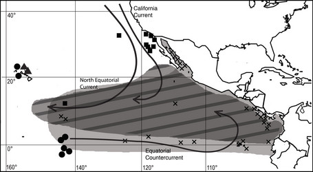
Figure. Distribution of pterygioteuthids in the tropical Pacific. Black circles = P. giardi. Black squares = P. gemmata. Crosses = P. microlampas. X = P. hoylei. Note- P. gemmata and P. giardi are also found in the Atlantic, while P. microlampas and P. hoylei are limited to the Pacific (modified from Lindgren, 2010).
The range of Pterygioteuthis hoylei beyond the ETP remains unclear. Records from Riddell (1985) and Nesis (1973) indicate that P. hoylei or a yet undescribed species may occur in southern waters off New Zealand and in the Indo-West Pacific.
References
Lindgren, A.R. 2010. Systematics and distribution of the squid genus Pterygioteuthis (Cephalopoda: Oegopsida) in the eastern tropical Pacific Ocean
Nesis, K.N. 1973. Cephalopods of the eastern equatorial and south-eastern Pacific Ocean (in Russian). In: English translations of selected publications on Cephalopods by Kir Nesis. Vol. I (M.J. Sweeney, compiler), pp. 171-248. Smithsonian Institution Libraries, Aad Publications, Karachi.
Okutani, T. 1974. Epipelagic decapod cephalopods collected by midwater tows during the EASTROPAC Expedition, 1967-1968 (systematic part). Bull. Tokai Reg. Fisheries Res. Lab. 80:29-118.
Pfeffer, G. 1912. Die Cephalopoden der Plankton-Expedition. Ergebnisse der Plankton-Expedition der Humboldt-Stiftung. 2: 1-815.
Riddell, D.J. 1985. Enoploteuthidae of the New Zealand Region. Fish. Res. Bull., New Zealand. 27:1-52.
Young, R.E. 1972. The systematics and areal distribution of pelagic cephalopods from the seas off southern California. Smith. Contr. Zool. 97:1-59.
Title Illustrations
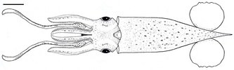
| Scientific Name | Pterygioteuthis hoylei |
|---|---|
| Specimen Condition | line drawing |
| Sex | Male |
| Life Cycle Stage | mature |
| View | dorsal |
| Copyright | © 2010 Journal of Molluscan Studies |
About This Page

Ohio State University, Columbus, Ohio, USA
University of Hawaii, Honolulu, HI, USA
Correspondence regarding this page should be directed to Annie Lindgren at and Richard E. Young at
Page copyright © 2011 and
All Rights Reserved.
- First online 11 January 2011
- Content changed 11 January 2011
Citing this page:
Lindgren, Annie and Richard E. Young. 2011. Pterygioteuthis hoylei previously considered Pterygioteuthis giardi hoylei. Version 11 January 2011 (under construction). http://tolweb.org/Pterygioteuthis_hoylei/68253/2011.01.11 in The Tree of Life Web Project, http://tolweb.org/


