


Pterygioteuthis giardi group
Annie Lindgren, Richard E. Young, and Katharina M. Mangold (1922-2003)


This tree diagram shows the relationships between several groups of organisms.
The root of the current tree connects the organisms featured in this tree to their containing group and the rest of the Tree of Life. The basal branching point in the tree represents the ancestor of the other groups in the tree. This ancestor diversified over time into several descendent subgroups, which are represented as internal nodes and terminal taxa to the right.
You can click on the root to travel down the Tree of Life all the way to the root of all Life, and you can click on the names of descendent subgroups to travel up the Tree of Life all the way to individual species.
For more information on ToL tree formatting, please see Interpreting the Tree or Classification. To learn more about phylogenetic trees, please visit our Phylogenetic Biology pages.
close boxIntroduction
Brief Diagnosis:
A Pterygioteuthis with...
- hooks in 2 series on arms I-III.
- tentacular club suckers on manus of similar size in transverse rows.
- eyes each with 10 large and 5 minute photophores.
Characteristics
- Arms - hooks are in 2 series on arms I-III.
Click on an image to view larger version & data in a new window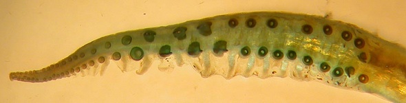
Figure. Oral view of arms I from a female P. hoylei. ©
- Arms I - distal suckers are globular.
Click on an image to view larger version & data in a new window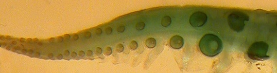
Figure. Close-up of distal suckers for P. hoylei. ©
- Arms IV - females without suckers or hooks, males with 2-3 hooks.
Click on an image to view larger version & data in a new window
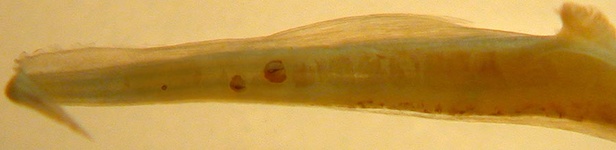
Figure. Oral views of right arms IV from P. hoylei. Top - female. Bottom - male with 2 hooks and 1 small sucker. ©
- Hectocotylus plate - contains 2 large teeth.
Click on an image to view larger version & data in a new window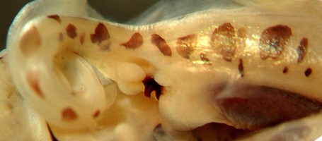
Figure. Hectocotylus of P. hoylei. ©
- Arms I - distal suckers are globular.
- Tentacular clubs - suckers (4 series) on manus of similar size in transverse rows.
Click on an image to view larger version & data in a new window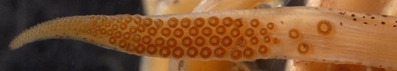
Figure. Oral view of the tentacular club from P. hoylei. ©
- Buccal membrane - with ridges on inner surface.
Click on an image to view larger version & data in a new window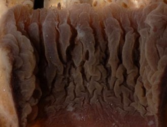
Click on an image to view larger version & data in a new windowFigure. Oral view of the buccal membrane from P. hoylei. ©
- Eye photophores - 10 large and 5 minute (see arrows below).
Click on an image to view larger version & data in a new window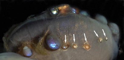
Figure. Ventral view of the minute eye photophores (arrows) from the right eye of P. giardi. ©
Comparison table for P. giardi and P. hoylei
| Character | P. giardi | P. hoylei |
| Arm I hooks | In 2 distinct sizes: regular and minute | Minute hooks absent |
| Male Arm III suckers | proximal suckers, distal hooks | hooks only* |
| Tentacular photophores | 2 | 4 |
| Tentacular stalk chromatophores | Stop midway up stalk (distal to 2nd photophore) | Continue along entire stalk in 1 row |
| Aboral tentacular club chromatophores | Absent | In 2 rows along edges of club |
| Funnel chromatophores | Few present anterior to funnel locking apparatus, generally absent from ventral funnel | Present anterior to locking apparatus, present on ventral funnel. |
Discussion of Phylogenetic Relationships
Pterygioteuthis hoylei was originally considered a subspecies of P. giardi: Hoyle (1904) was the first to note differences in 4 specimens of P. giardi collected from the eastern tropical Pacific, but he made no formal designation. Pfeffer (1912) later erected the subspecies, Pterygioteuthis giardi hoylei based on the descriptions by Hoyle, but made no further examination of specimens himself. Further investigation (Lindgren, 2010) uncovered notable differences between the two subspecies (see table above), leading to the elevation of P. hoylei to specific status.References
Lindgren, A.R. 2010. Systematics and distribution of the squid genus Pterygioteuthis (Cephalopoda: Oegopsida) in the eastern tropical Pacific Ocean
Nesis, K.N. 1973. Cephalopods of the eastern equatorial and south-eastern Pacific Ocean (in Russian). In: English translations of selected publications on Cephalopods by Kir Nesis. Vol. I (M.J. Sweeney, compiler), pp. 171-248. Smithsonian Institution Libraries, Aad Publications, Karachi.
Okutani, T. 1974. Epipelagic decapod cephalopods collected by midwater tows during the EASTROPAC Expedition, 1967-1968 (systematic part). Bull. Tokai Reg. Fisheries Res. Lab. 80:29-118.
Pfeffer, G. 1912. Die Cephalopoden der Plankton-Expedition. Ergebnisse der Plankton-Expedition der Humboldt-Stiftung. 2: 1-815.
Riddell, D.J. 1985. Enoploteuthidae of the New Zealand Region. Fish. Res. Bull., New Zealand. 27:1-52.
Young, R.E. 1972. The systematics and areal distribution of pelagic cephalopods from the seas off southern California. Smith. Contr. Zool. 97:1-59.
Title Illustrations

| Scientific Name | Pterygioteuthis |
|---|---|
| Specimen Condition | Dead Specimen |
| Identified By | S. Haddock |
| Life Cycle Stage | Juvenile |
| View | Ventral |
| Copyright | © 2006 Steven Haddock |
About This Page

Ohio State University, Columbus, Ohio, USA
University of Hawaii, Honolulu, HI, USA
Katharina M. Mangold (1922-2003)
Laboratoire Arago, Banyuls-Sur-Mer, France
Correspondence regarding this page should be directed to Annie Lindgren at and Richard E. Young at
Page copyright © 2011 and
 Page: Tree of Life
Pterygioteuthis giardi group.
Authored by
Annie Lindgren, Richard E. Young, and Katharina M. Mangold (1922-2003).
The TEXT of this page is licensed under the
Creative Commons Attribution-NonCommercial License - Version 3.0. Note that images and other media
featured on this page are each governed by their own license, and they may or may not be available
for reuse. Click on an image or a media link to access the media data window, which provides the
relevant licensing information. For the general terms and conditions of ToL material reuse and
redistribution, please see the Tree of Life Copyright
Policies.
Page: Tree of Life
Pterygioteuthis giardi group.
Authored by
Annie Lindgren, Richard E. Young, and Katharina M. Mangold (1922-2003).
The TEXT of this page is licensed under the
Creative Commons Attribution-NonCommercial License - Version 3.0. Note that images and other media
featured on this page are each governed by their own license, and they may or may not be available
for reuse. Click on an image or a media link to access the media data window, which provides the
relevant licensing information. For the general terms and conditions of ToL material reuse and
redistribution, please see the Tree of Life Copyright
Policies.
- First online 11 January 2011
- Content changed 11 January 2011
Citing this page:
Lindgren, Annie, Richard E. Young, and Katharina M. Mangold (1922-2003). 2011. Pterygioteuthis giardi group. Version 11 January 2011 (under construction). http://tolweb.org/Pterygioteuthis_giardi_group/110605/2011.01.11 in The Tree of Life Web Project, http://tolweb.org/

