Back to Profile Page

Tree of Life Media Contributed By Klaus Rohde

<< < 3 4 5 6 7 8 9 > >>
ID Thumbnail Media Data
2798
Reference redrawn from Rohde, K. and Watson, N. (1990a). Ultrastructural studies of juvenile Austramphilina elongata. Transmission electron-microscopy of sensory receptors. Parasitology Research 76, 336-342.
Life Cycle Stage juvenile
Body Part Sensory receptors
Image Use creative commons This media file is licensed under the Creative Commons Attribution-ShareAlike License - Version 3.0.
Copyright © Klaus Rohde
Attached to Group Gigantolina elongata: view page image collection
Title fig17am.gif
Image Type Diagram
Image Content Body Parts
Technical Information based on electron-microscopic studies
ID 2798
2826
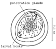
Comments Note lack of egg stalk. A side view, C apical view, B larval hooks (only two drawn).
Reference From Dubinina, M.N. (1982). Parasitic worms of the class Amphilinida (Platyhelminthes). "Nauka", Leningrad.
Acknowledgements According to Dönges and Harder (1966)
Life Cycle Stage egg, larva
Body Part larval hooks
Copyright © 1982 Nauka Publishers
Image Use ToL use only
Attached to Group Schizochoerus africanus: view page image collection
Title afri4.gif
Image Type Drawing/Painting
Image Content Specimen(s)
ID 2826
2850
Scientific Name Lobatostoma manteri
Reference redrawn from Rohde, K. and Watson, N. A. (1992). Sense receptors of larval Lobatostoma manteri (Trematoda, Aspidogastrea). International Journal for Parasitology 22, 35-42.
Life Cycle Stage larva
Body Part Sensory receptors
Image Use creative commons This media file is licensed under the Creative Commons Attribution-ShareAlike License - Version 3.0.
Copyright © Klaus Rohde
Attached to Group Aspidogastrea: view page image collection
Title fig13as.gif
Image Type Diagram
Image Content Body Parts
ID 2850
2979
Scientific Name Multicotyle purvisi
Reference redrawn from Rohde, K. (1972). The Aspidogastrea, especially Multicotyle purvisi Dawes, 1941. Advances in Parasitology 10, 77-151.
Life Cycle Stage adult
Body Part Note the septum of connective tissue between the anterior ventral and dorsal parts of the body
View Sagittal (longitudinal section along the midline)
Image Use creative commons This media file is licensed under the Creative Commons Attribution-ShareAlike License - Version 3.0.
Copyright © Klaus Rohde
Attached to Group Aspidogastrea: view page image collection
Title fig25as.gif
Image Type Drawing/Painting
Image Content Body Parts
ID 2979
3116
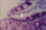
Scientific Name Peristernia australiensis
Comments Section through digestive gland (D) and stomach (S) of uninfected snail. Note: no thick fibrous layer below stomach epithelium of snail.
Specimen Condition Dead Specimen
Image Use creative commons This media file is licensed under the Creative Commons Attribution-ShareAlike License - Version 3.0.
Copyright © Klaus Rohde
Attached to Group Buccinoidea (Caenogastropoda): view page image collection
Title fig37as.gif
Image Type Photograph
Image Content Body Parts
ID 3116
3129
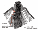
Reference Redrawn from Dubinina, M.N. (1982). Parasitic worms of the class Amphilinida (Platyhelminthes). "Nauka", Leningrad.
Acknowledgements According to Woodland (1923)
Body Part Anterior end, proboscis extended
Image Use creative commons This media file is licensed under the Creative Commons Attribution-ShareAlike License - Version 3.0.
Copyright © Klaus Rohde
Attached to Group Schizochoerus paragonopora: view page image collection
Title paragon2.gif
Image Type Drawing/Painting
Image Content Body Parts
ID 3129
3252
Reference redrawn from Dubinina, M.N. (1982). Parasitic worms of the class Amphilinida (Platyhelminthes). "Nauka", Leningrad.
Life Cycle Stage adult
Body Part vaginal opening and male gonopore, and various reproductive organs and ducts
Image Use creative commons This media file is licensed under the Creative Commons Attribution-ShareAlike License - Version 3.0.
Copyright © Klaus Rohde
Attached to Group Amphilina foliacea: view page image collection
Title fig15am.gif
Image Type Drawing/Painting
Image Content Body Parts
ID 3252
3295
Comments Note lack of egg stalk
Reference redrawn from Dubinina, M.N. (1982). Parasitic worms of the class Amphilinida (Platyhelminthes). "Nauka", Leningrad.
Life Cycle Stage Egg with larva
Image Use creative commons This media file is licensed under the Creative Commons Attribution-ShareAlike License - Version 3.0.
Copyright © Klaus Rohde
Attached to Group Gigantolina magna: view page image collection
Title magna4.gif
Image Type Drawing/Painting
Image Content Specimen(s)
ID 3295
3309
Scientific Name Multicotyle purvisi
Reference redrawn from Rohde, K. and Watson, N. (1991). Ultrastructure of pigmented photoreceptor of larval Multicotyle purvisi (Trematoda, Aspidogastrea). Parasitology Research 77, 485-490.
Life Cycle Stage larva
Body Part Ocellus (eye, photoreceptor)
Image Use creative commons This media file is licensed under the Creative Commons Attribution-ShareAlike License - Version 3.0.
Copyright © Klaus Rohde
Attached to Group Aspidogastrea: view page image collection
Title fig11as.gif
Image Type Diagram
Image Content Body Parts
ID 3309
3360
Body Part Note large sac-like accessory seminal receptacle at posterior end, and scattered frontal gland cells near anterior end.
Image Use creative commons This media file is licensed under the Creative Commons Attribution-ShareAlike License - Version 3.0.
Copyright © 2000 Klaus Rohde
Attached to Group Gigantolina elongata: view page image collection
Title elong1.gif
Image Type Drawing/Painting
Image Content Body Parts
ID 3360
Please note: Most images and other media displayed on the Tree of Life web site are protected by copyright, and the ToL cannot act as an agent for their distribution. If you would like to use any of these materials for your own projects, you need to ask the copyright owner(s) for permission. For additional information, please refer to the ToL Copyright Policies.

Contribute to the ToL

ToL Contributions

Ways to Contribute

Use of Contributions

Scientific Content

Building Treehouses

Images/Media

  go to the Tree of Life home page
top