Back to Profile Page

Tree of Life Media Contributed By Michel Laurin

<< < 1 2 3 4 5 6 7 > >>
ID Thumbnail Media Data
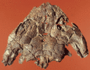
2140
Specimen Condition Fossil
Image Use creative commons This media file is licensed under the Creative Commons Attribution-NonCommercial License - Version 3.0.
Copyright © 1996 Michel Laurin
Attached to Group Utegenia shpinari: view page image collection
Title Utegenia_skull.gif
Image Type Photograph
Image Content Body Parts
ID 2140
2262
Image Use creative commons This media file is licensed under the Creative Commons Attribution-NonCommercial License - Version 3.0.
Copyright © 1996 Michel Laurin
Attached to Group Seymouriamorpha: view page image collection
Title Seymouriamorpha.gif
Image Type Drawing/Painting
Image Content Body Parts
ID 2262
2422
Comments Development of extraembryonic membranes in an amniote egg (chick). In this early developmental stage, the yolk sac is expanding over the yolk. The amnion and chorion are expanding over the embryo and will eventually form the amniotic chamber. The allantois is expanding toward the chorion, with which it will form a respiratory membrane, in addition to storing metabolic wastes of the embryo.
Reference Redrawn from Campbell, N. A. 1993. Biology. 3rd edition. New York: The Benjamin/Cummings Publishing.
Image Use creative commons This media file is licensed under the Creative Commons Attribution-NonCommercial License - Version 3.0.
Copyright © 1996 Michel Laurin
Attached to Group Amniota: view page image collection
Title Amniote_egg.gif
Image Type Drawing/Painting
Image Content Specimen(s), Body Parts
ID 2422
2492
Comments A, Cotylorhynchus, a Lower Permian synapsid; B, Captorhinus, a Lower Permian romeriid; C, Procolophon, a Triassic anapsid; D, Proganochelys, the oldest known turtle (Upper Triassic). Redrawn from A, Laurin & Reisz (1995); B, Heaton (1979); C, Carroll & Lindsay (1985); and D, Gaffney (1990). Abbreviations: Bc, braincase; E, epipterygoid; F, frontal; L, lacrimal; M, maxilla; N, nasal; P, parietal; Pm, premaxilla; Po, postorbital; Pof, postfrontal; Prf, prefrontal; Pt, pterygoid; Q, quadrate; Qj, quadratojugal; Sq, squamosal; St, supratemporal; T, tabular.
Body Part skulls
View lateral
Image Use creative commons This media file is licensed under the Creative Commons Attribution-NonCommercial License - Version 3.0.
Copyright © 1996 Michel Laurin
Attached to Group Amniota: view page image collection
Title Amniota_lat.gif
Image Type Drawing/Painting
Image Content Body Parts
ID 2492
2727
Scientific Name Spinoaequalis schultzei, Haptodus garnettensis, Sigillaria
Comments The Pennsylvanian diapsid Spinoaequalis schultzei jumps into the water to escape a hungry Haptodus garnettensis (a synapsid). The base of a Sigillaria trunk is visible in the upper right corner.
Creator Michael deBraga
Copyright © 2000 Michael deBraga
Image Use restricted
Attached to Group Diapsida: view page image collection
Title spinoaequalis.gif
Image Type Drawing/Painting
Image Content Specimen(s)
ID 2727
2864
Location Texas
Reference Reproduced from White, T. E. 1939. Osteology of Seymouria baylorensis Broili. Bulletin of the Museum of Comparative Zoology 85:325-409.
Specimen Condition Fossil -- Period: Lower Permian
Copyright © 1939 The President and Fellows of Harvard College
Image Use restricted
Attached to Group Seymouria baylorensis (Seymouria): view page image collection
Title s.baylorensis.gif
Image Type Drawing/Painting
Image Content Specimen(s)
ID 2864
3029
Location Germany
Comments Restored environment of Proganochelys (the oldest known turtle) during the Late Triassic in Germany. Besides Proganochelys, small hybodont sharks are shown.
Creator Painting by Frank Ippolito
Specimen Condition Fossil -- Period: Late Triassic
Copyright © 1987 American Museum of Natural History
Image Use restricted
Attached to Group Proganochelys (Testudines): view page image collection
Title Proganochelys.gif
Image Type Drawing/Painting
Image Content Specimen(s)
ID 3029
3275
Image Use creative commons This media file is licensed under the Creative Commons Attribution-NonCommercial License - Version 3.0.
Copyright © 2000 Michel Laurin
Attached to Group Diapsida: view page image collection
Title diapsida_pal.gif
Image Type Drawing/Painting
Image Content Body Parts
ID 3275
3679
Image Use creative commons This media file is licensed under the Creative Commons Attribution-NonCommercial License - Version 3.0.
Copyright © 2000 Michel Laurin
Attached to Group Diapsida: view page image collection
Title diapsida_dor.gif
Image Type Drawing/Painting
Image Content Body Parts
ID 3679
3757
Comments The photograph shows the dermal sculpturing that characterizes relatively mature seymouriamorphs.
Specimen Condition Fossil
Life Cycle Stage juvenile postmetamorphic
Body Part skull
View dorsal
Image Use creative commons This media file is licensed under the Creative Commons Attribution License - Version 3.0.
Copyright ©
Attached to Group Discosauriscus austriacus (Discosauriscus): view page image collection
Title Disco_juve.gif
Image Type Photograph
Image Content Body Parts
ID 3757
Please note: Most images and other media displayed on the Tree of Life web site are protected by copyright, and the ToL cannot act as an agent for their distribution. If you would like to use any of these materials for your own projects, you need to ask the copyright owner(s) for permission. For additional information, please refer to the ToL Copyright Policies.

Contribute to the ToL

ToL Contributions

Ways to Contribute

Use of Contributions

Scientific Content

Building Treehouses

Images/Media

  go to the Tree of Life home page
top