|
ID
|
Thumbnail |
Media Data |
| 30363 |
|
|
Comments
|
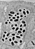
infected human enterocyte
|
|
Specimen Condition
|
Dead Specimen
|
|
Life Cycle Stage
|
developmental stages and spores
|
|
Image Use
|
 This media file is licensed under the Creative Commons Attribution-NonCommercial License - Version 3.0. This media file is licensed under the Creative Commons Attribution-NonCommercial License - Version 3.0.
|
|
Copyright
|
© J.M. Orenstein
|
|
Attached to Group
|
Encephalitozoon intestinalis (Microsporidia): view page image collection
|
|
Title
|
septata1.jpg
|
|
Image Type
|
Photograph
|
|
Image Content
|
Specimen(s), Ultrastructure
|
|
Technical Information
|
transmission electron micrograph
|
|
ID
|
30363
|
|
| 30364 |
|
|
Specimen Condition
|
Dead Specimen
|
|
Life Cycle Stage
|
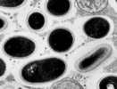
spores
|
|
Body Part
|
parasitophorous vacuole showing imperfect septa
|
|
Image Use
|
This media file is licensed under the Creative Commons Attribution-NonCommercial License - Version 3.0.
|
|
Copyright
|
© J.M. Orenstein
|
|
Attached to Group
|
Encephalitozoon intestinalis (Microsporidia): view page image collection
|
|
Title
|
septata2.jpg
|
|
Image Type
|
Photograph
|
|
Image Content
|
Specimen(s), Ultrastructure
|
|
Technical Information
|
Transmission electron micrograph
|
|
ID
|
30364
|
|
| 30365 |
|
|
| 31008 |
|
|
Scientific Name
|
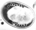
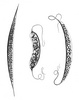
Caudospora sp.
|
|
Comments
|
fresh spore isolated from the fat body of Prosimulium mixtum (Diptera: Simuliidae). The spores of aquatic microsporidia bear often surface ornamentations, probably serving as floating devices.
|
|
Specimen Condition
|
Live Specimen
|
|
Life Cycle Stage
|
spore
|
|
Copyright
|
© 1979

|
|
Image Use
|
ToL use only
|
|
Attached to Group
|
Caudospora (Microsporidia): view page image collection
|
|
Title
|
caudospora.jpg
|
|
Image Type
|
Photograph
|
|
Image Content
|
Specimen(s)
|
|
ID
|
31008
|
|
| 30990 |
|
|
Comments
|
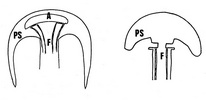
Spore apex at typical microsporidia (left) vs. Metchnikovella-like microsporidia (right). PS- polar sac, A- anchoric disc, F- polar filament.
|
|
Life Cycle Stage
|
spore
|
|
Body Part
|
Spore apex
|
|
Copyright
|
© xx xx
|
|
Image Use
|
ToL use only
|
|
Attached to Group
|
typical microsporidia (Microsporidia): view page image collection
Metchnikovella-like microsporidia (Microsporidia): view page image collection
|
|
Title
|
polarsac2_typical_vs_atypical.jpg
|
|
Image Type
|
Diagram
|
|
Image Content
|
Body Parts, Ultrastructure
|
|
Notes
|
KS: Where does this image come from? Who is the copyright owner? We cannot release the full-size image until copyright ownership has been resolved.
|
|
ID
|
30990
|
|
| 30992 |
|
|
Scientific Name
|
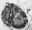
Amphiacantha longa
|
|
Reference
|
Larsson J. I. R. 2000. The hyperparasitic microsporidium Amphiacantha longa Caullery et Mesnil, 1914 (Microspora: Metchnikovellidae) description of the cytology, redescription of the species, emended diagnosis of the genus Amphiacantha and establishment of the new family Amphiacanthidae. Folia Parasitologica 47: 241-256.
|
|
Specimen Condition
|
Dead Specimen
|
|
Life Cycle Stage
|
spore
|
|
Body Part
|
A-bulbous swelling-appendix, C-circular element, EX-exospore, ER-endoplasmic reticulum, F-polar filament-manubrium, MC-membranous coat, N-nucleus
|
|
Copyright
|
© 2000 Institute of Parasitology, Academy of Sciences of the Czech Republic, České Budějovice
|
|
Image Use
|
ToL use only
|
|
Attached to Group
|
Amphiacantha (Microsporidia): view page image collection
|
|
Title
|
amphiacadobe.jpg
|
|
Image Type
|
Photograph
|
|
Image Content
|
Specimen(s), Ultrastructure
|
|
Technical Information
|
TEM micrograph
|
|
Notes
|
permission pending
|
|
ID
|
30992
|
|
| 30993 |

|
|
Scientific Name
|
Amphiacantha longa and Lecudina
|
|
Comments
|
in the cell of the gregarine Lecudina (gregarine nucleus is at *)
|
|
Reference
|
Larsson J. I. R. 2000. The hyperparasitic microsporidium Amphiacantha longa Caullery et Mesnil, 1914 (Microspora: Metchnikovellidae) description of the cytology, redescription of the species, emended diagnosis of the genus Amphiacantha and establishment of the new family Amphiacanthidae. Folia Parasitologica 47: 241-256.
|
|
Specimen Condition
|
Live Specimen
|
|
Life Cycle Stage
|
spore sacs (arrow) and free spores (arrowheads)
|
|
Copyright
|
© 2000 Institute of Parasitology, Academy of Sciences of the Czech Republic, České Budějovice
|
|
Image Use
|
ToL use only
|
|
Attached to Group
|
Lecudinidae (Gregarina): view page image collection
Amphiacantha (Microsporidia): view page image collection
|
|
Title
|
amphiacantha.jpg
|
|
Image Type
|
Photograph
|
|
Image Content
|
Specimen(s), Body Parts
|
|
Notes
|
permission pending
|
|
ID
|
30993
|
|
| 30994 |
|
|
Scientific Name
|
Amphiacantha longa, Amphiacantha ovalis, Amphiacantha attenuata
|
|
Reference
|
Vivier E. 1975. The microsporidia of the Protozoa. Protistologica 11: 345-361.
|
|
Life Cycle Stage
|
spore sacs and spores
|
|
Copyright
|
© 1975 Éditions du Centre national de la recherche scientifique
|
|
Image Use
|
ToL use only
|
|
Attached to Group
|
Amphiacantha (Microsporidia): view page image collection
|
|
Title
|
amphiacanthaschema.jpg
|
|
Image Type
|
Drawing/Painting
|
|
Image Content
|
Specimen(s), Body Parts
|
|
ID
|
30994
|
|
| 30995 |

|
|
Scientific Name
|
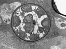
Chytridiopsis typographi
|
|
Specimen Condition
|
Dead Specimen
|
|
Life Cycle Stage
|
spore
|
|
Copyright
|
© 2008
|
|
Image Use
|
restricted
|
|
Attached to Group
|
Chytridiopsis (Microsporidia): view page image collection
|
|
Title
|
chytrdsporaexcelentni.jpg
|
|
Image Type
|
Photograph
|
|
Image Content
|
Specimen(s), Ultrastructure
|
|
Technical Information
|
TEM micrograph
|
|
ID
|
30995
|
|
| 30996 |
|
|
Scientific Name
|
Chytridiopsis typographi
|
|
Specimen Condition
|
Dead Specimen
|
|
Life Cycle Stage
|
thickwalled spore sac with young spores
|
|
Copyright
|
© 2008
|
|
Image Use
|
restricted
|
|
Attached to Group
|
Chytridiopsis (Microsporidia): view page image collection
|
|
Title
|
chytridcystalepsi.jpg
|
|
Image Type
|
Photograph
|
|
Image Content
|
Specimen(s), Ultrastructure
|
|
Technical Information
|
TEM micrograph
|
|
ID
|
30996
|
|
