|
ID
|
Thumbnail |
Media Data |
| 1662 |

|
|
Comments
|
Note the ciliated epidermis overlying the tegument (neodermis) with its basal lamina. The perikarya (nucleated parts of the cells) of the tegument are "insunk", i.e., they are located deep in the tissue. The ciliated epidermis is shed when the larva penetrates into a crayfish, and the tegument becomes the surface layer of the juvenile and adult
|
|
Reference
|
redrawn from Rohde, K. and Georgi, M. (1983). Structure and development of the larva of Austramphilina elongata Johnston, 1931 (Cestodaria, Amphilinidea). International Journal for Parasitology 13, 273-287.
|
|
Life Cycle Stage
|
larva
|
|
View
|
Longitudinal section through surface layers
|
|
Image Use
|
 This media file is licensed under the Creative Commons Attribution-ShareAlike License - Version 3.0. This media file is licensed under the Creative Commons Attribution-ShareAlike License - Version 3.0.
|
|
Copyright
|
© Klaus Rohde

|
|
Attached to Group
|
Gigantolina elongata: view page image collection
|
|
Title
|
fig10am.gif
|
|
Image Type
|
Diagram
|
|
Image Content
|
Body Parts
|
|
Technical Information
|
based on electron-microscopic studies
|
|
ID
|
1662
|
|
| 1702 |
|
|
Comments
|
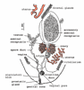
Note separate vaginal and male pores, deeply lobed ovary consisting of two wings, and large accessory seminal receptacle in addition to seminal receptacle.
|
|
Reference
|
redrawn from Dubinina, M.N. (1982). Parasitic worms of the class Amphilinida (Platyhelminthes). "Nauka", Leningrad.
|
|
Body Part
|
Posterior end
|
|
Image Use
|
This media file is licensed under the Creative Commons Attribution-ShareAlike License - Version 3.0.
|
|
Copyright
|
© Klaus Rohde
|
|
Attached to Group
|
Gigantolina magna: view page image collection
|
|
Title
|
magna3.gif
|
|
Image Type
|
Drawing/Painting
|
|
Image Content
|
Body Parts
|
|
ID
|
1702
|
|
| 1769 |
|
|
Scientific Name
|
Multicotyle purvisi
|
|
Reference
|
redrawn from Rohde, K. (1972). The Aspidogastrea, especially Multicotyle purvisi Dawes, 1941. Advances in Parasitology 10, 77-151.
|
|
Life Cycle Stage
|
larva
|
|
Body Part
|
Note four anterior and six posterior ciliary tufts
|
|
Image Use
|
This media file is licensed under the Creative Commons Attribution-ShareAlike License - Version 3.0.
|
|
Copyright
|
© Klaus Rohde
|
|
Attached to Group
|
Aspidogastrea: view page image collection
|
|
Title
|
fig05as.gif
|
|
Image Type
|
Diagram
|
|
Image Content
|
Specimen(s)
|
|
ID
|
1769
|
|
| 1830 |

|
|
Comments
|
Note elongate accessory seminal receptacle.
|
|
Reference
|
Redrawn from Dubinina, M.N. (1982). Parasitic worms of the class Amphilinida (Platyhelminthes). "Nauka", Leningrad.
|
|
Body Part
|
posterior end
|
|
Image Use
|
This media file is licensed under the Creative Commons Attribution-ShareAlike License - Version 3.0.
|
|
Copyright
|
© Klaus Rohde
|
|
Attached to Group
|
Schizochoerus liguloideus: view page image collection
|
|
Title
|
ligul2.gif
|
|
Image Type
|
Drawing/Painting
|
|
Image Content
|
Body Parts
|
|
ID
|
1830
|
|
| 1890 |

|
|
Image Use
|
restricted
|
|
Title
|
dot.gif
|
|
Image Type
|
Photograph
|
|
Image Content
|
Specimen(s)
|
|
ID
|
1890
|
|
| 1913 |
|
|
Scientific Name
|
Lobatostoma manteri
|
|
Comments
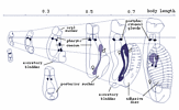
|
Development after hatching. Note the transformation of the posterior sucker into the ventral disc, and - connected with this - the change in the body proportions
|
|
Reference
|
redrawn from Rohde, K. (1975). Early development and pathogenesis of Lobatostoma manteri Rohde (Trematoda: Aspidogastrea). International Journal for Parasitology 5, 597-607.
|
|
Image Use
|
This media file is licensed under the Creative Commons Attribution-ShareAlike License - Version 3.0.
|
|
Copyright
|
© Klaus Rohde
|
|
Attached to Group
|
Aspidogastrea: view page image collection
|
|
Title
|
fig17as.gif
|
|
Image Type
|
Diagram
|
|
Image Content
|
Specimen(s)
|
|
ID
|
1913
|
|
| 1968 |

|
|
| 2002 |

|
|
Comments
|
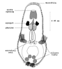
a. Arrangement of hooks in the larva, b. Shape of hooks in greater detail
|
|
Reference
|
redrawn from Dubinina, M.N. (1982). Parasitic worms of the class Amphilinida (Platyhelminthes). "Nauka", Leningrad.
|
|
Life Cycle Stage
|
larva
|
|
Body Part
|
Hooks
|
|
Image Use
|
This media file is licensed under the Creative Commons Attribution-ShareAlike License - Version 3.0.
|
|
Copyright
|
© Klaus Rohde
|
|
Attached to Group
|
Amphilina foliacea: view page image collection
|
|
Title
|
fig6am.gif
|
|
Image Type
|
Diagram
|
|
Image Content
|
Body Parts
|
|
ID
|
2002
|
|
| 2013 |
|
|
Reference
|
redrawn from Rohde, K. (1972). The Aspidogastrea, especially Multicotyle purvisi Dawes, 1941. Advances in Parasitology 10, 77-151.
|
|
Body Part
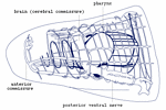
|
Nervous system in anterior part of body. Note that the brain is the dorsal part of a ring commissure, and that there is a second more external ring commissure at the same level
|
|
Image Use
|
This media file is licensed under the Creative Commons Attribution-ShareAlike License - Version 3.0.
|
|
Copyright
|
© Klaus Rohde
|
|
Attached to Group
|
Aspidogastrea: view page image collection
|
|
Title
|
fig30as.gif
|
|
Image Type
|
Drawing/Painting
|
|
Image Content
|
Body Parts
|
|
ID
|
2013
|
|
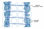
| 2079 |
|
|
Scientific Name
|
Multicotyle purvisi
|
|
Reference
|
redrawn from Rohde, K. (1972). The Aspidogastrea, especially Multicotyle purvisi Dawes, 1941. Advances in Parasitology 10, 77-151.
|
|
Body Part
|
marginal organs, showing marginal glands, ampullae and connecting ducts
|
|
Image Use
|
This media file is licensed under the Creative Commons Attribution-ShareAlike License - Version 3.0.
|
|
Copyright
|
© Klaus Rohde
|
|
Attached to Group
|
Aspidogastrea: view page image collection
|
|
Title
|
fig29as.gif
|
|
Image Type
|
Drawing/Painting
|
|
Image Content
|
Body Parts
|
|
ID
|
2079
|
|
