Drawings below are first made from photographs that are then compared with the preserved squid under a microscope. A small error will occur in identifying the two classes of photophores (red - counterilluminating photophores with red color filters = "complex photophores, and blue - simple photophores plus lens bearing photophores = "non-photophores." Blue photophores cannot, yet, be reliably separated, in preserved squid, into the two component types.). A damaged or developing/transforming photophore cannot always be reliably identified. Even the presence or absence of photophores can be difficult on strongly curved surfaces, for example, the sides of the head or mantle when viewed ventrally. Therefore, the photophore arrangement, seen below, is most accurate on the ventral surfaces when viewed ventrally and on the lateral surfaces when viewed laterally.

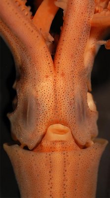
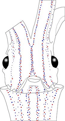
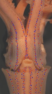
Figure. Ventral views of the integumental photophores of Abraliopsis sp. A, mature male, 30 mm ML, Hawaiian waters. Left - Photograph of the preserved squid. Middle - Outline drawing from photograph with all integumental photophores represented by colored dots. Right - Photograph with colored dots superimposed on integumental photophores. Red dots - Complex photophores. Blue dots - Non-complex photophores. Images by R. Young.
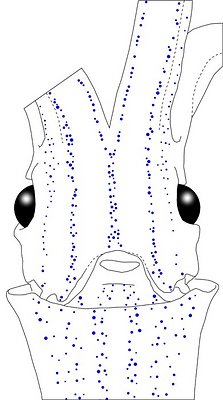
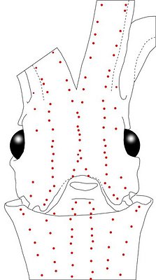
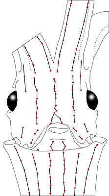
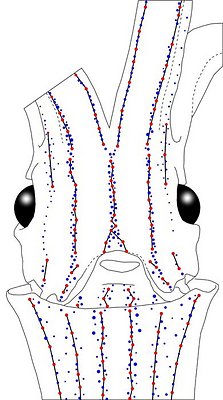
Figure. Same squid (30 mm ML), same view as above.. Top left - Drawing showing only the non-complex (blue) photophores. Top middle - Drawing showing only the complex (red) photophores . Top right - Same drawing with lines connecting red photophores to aid comparisons of photophore patterns between species. Bottom - Drawing showing all photophores and lines showing red photophore patterns. Images by R. Young.
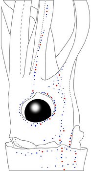
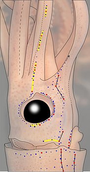
Figure. Side view of the same squid showing lateral photophores. Left - Photograph of the preserved squid. Middle - Outline drawing from photograph with all integumental photophores represented by colored dots. Right - Outline drawing over dimmed photograph with all integumental photophores represented by colored dots, and with lines connecting some photophores. Red dots - Complex photophores. Blue dots - Non-complex photophores. Lines connecting photophores assist in understanding the organization of the photophores. Black lines - Patterns based on red photophores. Yellow line - Pattern based on red and blue photophores. Images by R. Young.
Mature male, 32 mm ML
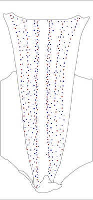

Figure. Ventral views of the mantle integumental photophores of Abraliopsis sp. A, mature male, 32 mm ML, Hawaiian waters. Left - Photograph of the preserved squid. Middle - Outline drawing from the photograph with all integumental photophores represented by colored dots. Right - Photograph (dimmed) with colored dots superimposed on integumental photophores. Red dots - Complex photophores. Blue dots - Non-complex photophores. Images by R. Young.
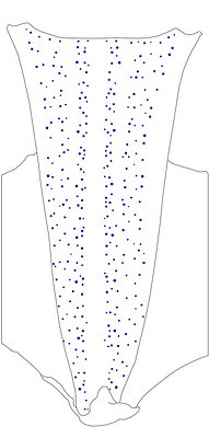
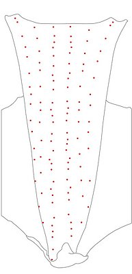
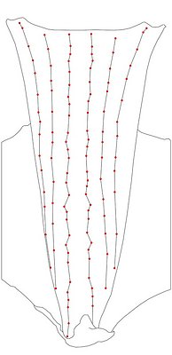
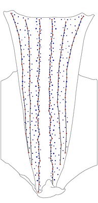
Figure. Same view, same squid. Left - Drawing showing only the non-complex (blue) photophores. Left middle - Drawing showing only the complex (red) photophores. Right middle - Same drawing with lines connecting red photophores added to aid comparisons of photophore patterns between species. Right - Drawing showing all photophores and lines showing red photophore patterns. Images by R. Young.
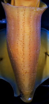
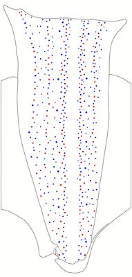
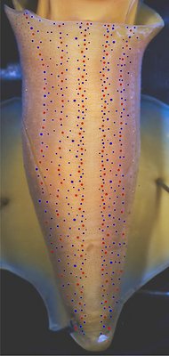
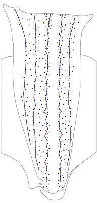
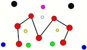
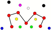
Figure. Ventral views of a larger female, Hawaiian waters. Left - Drawing showing only the non-complex (blue) photophores. Left middle - Drawing showing only the complex (red) photophores. Right middle - Same drawing with lines connecting red photophores added to aid comparisons of photophore patterns between species. Right - Drawing showing all photophores and lines showing red photophore patterns. Images by R. Young.
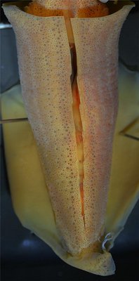
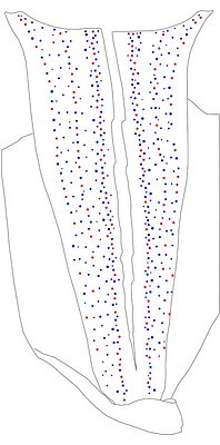
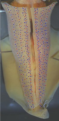
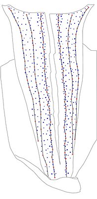
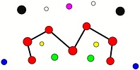
Figure. Ventral views of a larger male, Hawaiian waters. Left - Drawing showing only the non-complex (blue) photophores. Left middle - Drawing showing only the complex (red) photophores. Right middle - Same drawing with lines connecting red photophores added to aid comparisons of photophore patterns between species. Right - Drawing showing all photophores and lines showing red photophore patterns. Images by R. Young.
Comments:
The mantle photophores of the three squid illustrated here show the consistently large Medial Mantle Sector with blue photophores aligning close to the lines of red photophores on either side (ie, the Medial Mantle Series). Also, the Lateral Mantle Sectors show an absence of photophores in the midline of these sectors in about the anterior third of the mantle. This feature is more obvious in the 32 mm ML squid, which, because of its smaller size, has fewer overall photophores. The pattern, however, holds in the two larger squid.
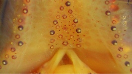
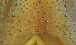
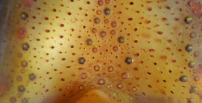
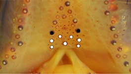
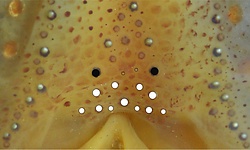
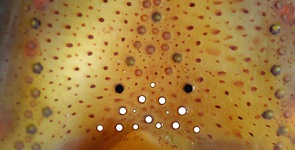
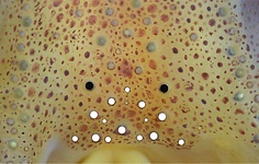
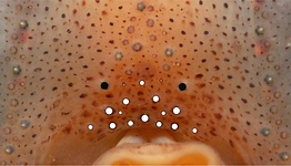
Tables. Ventral view of the funnel-groove region of 6 individuals of Abraliopsis sp, A. Top tier - Funnel groove showing photophores of funnel groove and surrounding tissue. Middle tier - In the photograph, white dots placed over the "white" photophores of the funnel groove. Black dots placed over the two most posteromedial "red" photophores of the ventral head to mark the anterolateral edges of the funnel groove. Bottom tier - Same pattern of dots as in photographs but the dots are colored. Colored dots, lines - Aid in comparison of patterns between species. Open dots - Represent photophores that are either of uncertain status as "white" photophores or have variable presence.
Note the ontogenetic effects. Images by R. Young. Comparison of white photophore patterns for all Abraliopsis species can be found here.
| Immature male, 18 mm ML | Immature male, 24 mm ML | Mature male, 28 mm ML |
|---|---|---|
 |  |  |
 |  |  |
 |  |  |
| Mature male, 31 mm ML | Mature male, 37 mm ML | Mature female, 47 mm ML |
|---|---|---|
 |  |  |
 |  |  |
 |  |  |
Comments: Summary of photophore distributions. (photophore definitions found here)
ARMS:
Medial Arm IV Series - Continuous; reaches about half of arm length.
Central Arm IV Sector - Photophores absent.
Lateral Arm IV Series - Discontinuous, extends to arm tip.
Lateral Membrane Series of arms IV - Reaches at least 70% of ML.
Arm III Series - Discontinuous; extends nearly to arm tip.
HEAD - Typical three photophore series on central-ventral head with:
Median Head Series - At least 5 "red" photophores in posterior caret. Anteriorly red photophores in irregular single series.
Median Head Sector - Absent.
Lateral Head Sector - Without photophores except for a few blue photophores in large squid.
Lateral Head Series - Single, nearly straight, series of red photophores.
Window Sector - Few, scattered, blue photophores anterolaterally, otherwise photophores absent.
1st Lateral Window Series - Two segments broadly separated by mostly-single series of blue photophores: posterior segment with 2 red photophores; anterior segment with two red photophore but extending onto lateral arm IV membrane with an additional red.
2nd Lateral Window Series - Absent.
Eyelid Series - Red photophores only on ventral half of eyelid. Posterior Eyelid Series with two large blue photophores displaced slightly posteriorly from eyelid series; more ventral of two, larger and more posterior. Formula: RbbBbB.
Occipital Series - Rbb.
Lateral Funnel-groove Series - 4 red photophores, in two segments not aligned.
White Patch (funnel groove) - Complex-2 Pattern (red, green, blue; + yellow, maroon at larger ML).
FUNNEL:
Medial Patch - "Nose" pattern of 4 "red" photophores.
Lateral Patch - Two red photophores.
MANTLE - Typical 6 photophore series on ventral mantle with:
Median Mantle Sector - Broad and without central photophores (bare) along entire mantle length. Blue photophores closely aligned to sector margins.
Medial Mantle Series - Nearly straight alignment in anterior half of mantle.
1st Lateral Mantle Sector - Scattered blue photophores present along margins of sector; bare strip extends down center of sector in anterior half of mantle. Extent of bare strip decreases with squid size to about one fourth of ML by 37 mm ML; in largest squid examined (47 mm ML) bare strip not recognizable.
2nd Lateral Mantle Sector - Sparse blue photophores.
Lateral Mantle and Mantle-angle Series - Both with virtually straight alignment (alignment anteriorly veers laterally as mantle broadens).
Comments
Within the subgenus Pfefferiteuthis, photophore patterns in Abraliopsis sp. A are especially similar to those of A. falco and Abraliopsis sp. P. In A. falco the median mantle sector has a slender bare strip but this is very broad the the other two species. In A. falco the first lateral mantle sector is densely packed with blue photophores but less so in the other two species.
The photophore series of A. atlantica and A. chuni are similar to the above but not well enough known to detect differences.


