Drawings below are first made from photographs that are then compared with the preserved squid under a microscope. A small error will occur in identifying the two classes of photophores (red - counterilluminating photophores with red color filters = "complex photophores, and blue - simple photophores plus lens bearing photophores = "non-complex photophores." Blue photophores cannot, yet, be reliably separated, in preserved squid, into the two component types.). A damaged or developing/transforming cannot always be reliably identified. Even the presence or absence of photophores can be difficult on strongly curved surfaces, for example, the sides of the head or mantle when viewed ventrally. Therefore, the photophore arrangement, seen below, is most accurate on the ventral surfaces when viewed ventrally and on the lateral surfaces when viewed laterally.

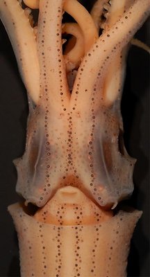
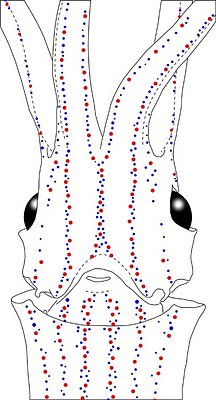
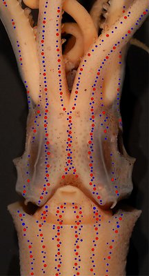
Figure. Ventral views of the integumental photophores of Abraliopsis sp. E, male, 21 mm ML, central equatorial Pacific. Left - Photograph of the preserved squid. Middle - Outline drawing with all integumental photophores represented by colored dots. Right - Photograph withcolored dots superimposed on integumental photophores. Red dots - Complex photophores. Blue dots - Non-complex photophores. Images by R. Young.
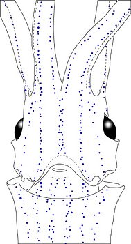
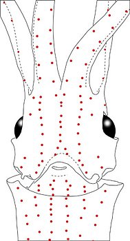
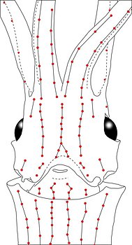
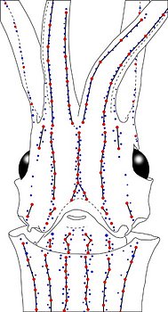
Figure. Same squid, same view as above. Left - Drawing showing only the non-complex (blue) photophores. Left middle - Drawing showing only the complex (red) photophores. Right middle - Same as previous drawing with lines connecting red photophores to aid comparisons of photophore patterns between species. Right - Drawing showing all photophores and lines showing red photophore patterns. Images by R. Young.
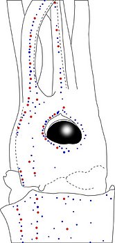
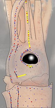
Figure. Side view of the head and proximal regions of the arms of the same squid (21 mm ML) as above. Left - Photograph of the preserved squid. Middle - Outline drawing from photograph with all integumental photophores represented by colored dots. Right - Outline drawing over dimmed photograph with all integumental photophores represented by colored dots, and with lines connecting some photophores. Red dots - Complex photophores. Blue dots - Non-complex photophores. Lines connecting photophores assist in understanding the organization of the photophores. Black lines - Patterns based on red photophores. Yellow line - Pattern based on red and blue photophores. Images by R. Young.
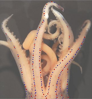
Figure. Ventral view of the integumental photophores of arms III and IV of Abraliopsis sp. E. Left - Photograph of the preserved squid. Right - Photograph dimmed with Colored dots placed over photophores to, more clearly, show the photophore distributions. Red dots - Complex photophores. Blue dots - Non-complex photophores.
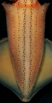
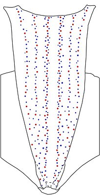
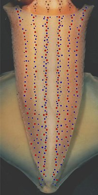
Figure. Ventral views of the integumental photophores of the same squid (21 mm ML). Left - Photograph of the preserved squid. Middle - Outline drawing from photograph with all integumental photophores represented by colored dots. Right - Photograph (dimmed) with colored dots superimposed on integumental photophores. Red dots - Complex photophores. Blue dots - Non-complex photophores. Images by R. Young.
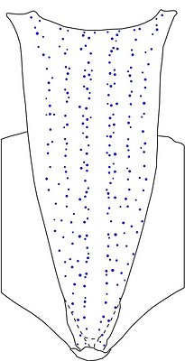
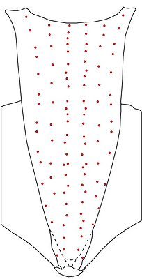
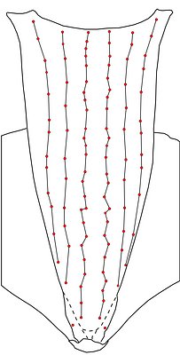
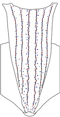
Figure. Ventral views of the mantle integumental photophores of the same squid. Left - Drawing showing only the non-complex (blue) photophores. Left-middle - Drawing showing only the complex (red) photophores. Right-middle - Same drawing with lines connecting red photophores to aid comparisons of photophore patterns between species. Right - Drawing showing all photophores, and lines showing red photophore patterns. Images by R. Young.
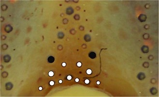
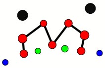
Figure. Ventral view of the funnel-groove region of Abraliopsis sp. E. Top - Photograph of the preserved squid showing photophores of the funnel groove and surrounding tissue. Middle - In the photograph, white dots are placed over the "white" photophores of the funnel groove. Black dots are placed over the two most posteromedial "red" photophores of the ventral head to mark the anterolateral edges of the funnel groove. Bottom - Same pattern of dots as in photographs but dots are colored. Colored dots, lines - Aid in comparison of patterns between species. Open dots - Represent photophores that are either of uncertain status as "white" photophores or have variable presence. Images by R. Young. Comparison of white photophore patterns for all Abraliopsis species can be found here.
Comments: Summary of photophore distributions.(photophore definitions found here)
ARMS:
Medial Arm IV Series - Continuous; reaches slightly less than 50 % of arm length.
Central Arm Sector - Photophores absent.
Lateral Arm IV Series - Nearly continuous (short breaks in distal half), reaches arm tip.
Lateral Membrane Series of arms IV - Reaches about 15% of arm length.
Arm III series - Discontinuous, reaches nearly 80% of arm length.
HEAD - Typical three photophore series on central-ventral head with:
Median Head Series - 5 "red" photophores in posterior caret. Anteriorly red photophores in regular single series.
Median Head Sector - Absent.
Lateral Head Sector - Photophores absent.
Window Sector - 2 blue photophores anterolaterally, otherwise photophores absent.
1st Lateral Window Series -Two segments broadly separated by single series of blue photophores: Both segments with 2 red photophores.
2nd Lateral Window Series - Absent.
Eyelid Series - Encircles eye opening with red photophores only on ventral half. Posterior Eyelid Series with two large blue photophores displaced slightly posteriorly from eyelid series; more ventral of two, larger and more posterior. Formula: RbbBbB.
Occipital Series - Rb.
Lateral Funnel-groove Series - 3 (1+2) red photophores.
White patch (funnel groove) - Complex-1 Pattern (red, green, blue).
FUNNEL:
Medial patch - "Nose" pattern of 4 "red" photophores.
Lateral patch - Two red photophores.
MANTLE - Typical 6 photophore series on ventral mantle with:
Median Mantle Sector - Broad; without photophores in central region over entire mantle length. Blue photophores closely aligned to sector margins.
Medial Mantle Series - Fairly well aligned in anterior half of mantle.
Lateral Mantle Sectors - In anterior half of mantle, few blue photophores, all located near margins in both sectors.
Lateral and Mantle-angle Series - Both with virtually straight alignment (alignment anteriorly veers laterally as mantle broadens).


