
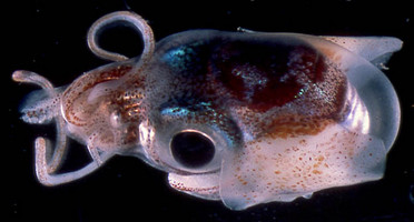
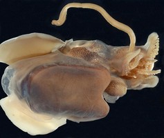
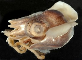
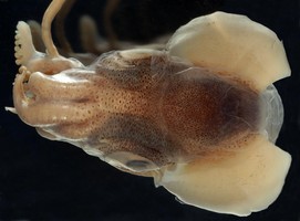
Figure. Dorsolateral view of I. iris. Photograph by Thomas Burch.
- Arms (Harman and Seki, 1990)
- Males: Each arm I with 20-28 suckers (mean 24.4); each arm II with 22-31 suckers (mean 25.1); each arm III with 24-31 suckers (mean 27.1); proximal half of each arm IV with 10-20 relatively large suckers, distal half with 70+ smmall suckers.
- Females: Each arm I with 18-32 suckers (mean 28.3); each arm II with 24-45 suckers (mean 39.0); each arm III with 18-27 suckers (mean 22.0); each arm IV with proximal half with 6-14 relatively large suckers in proximal half and next quarter with 10-30 smaller suckers.
- Arm bases proximal to suckers, in females, covered with small flattened papillae.
- Tentacles (Harman and Seki, 1990)
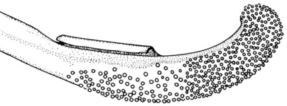
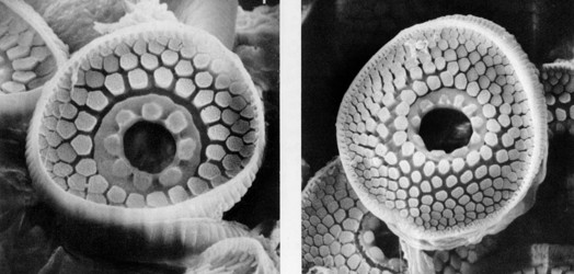
- Tentacular club with two sucker fields: distal half with minute, crowded suckers (ca. 19 suckers across), proximal suckers slightly larger, less crowded (ca. 11 across).
 Click on an image to view larger version & data in a new window
Figure. Oral view of the tentacular club of I. iris. Arrow points to tentacle organ. Note sucker sizes. Blue color from methylene blue stain. Photograph by R. Young.
- Funnel
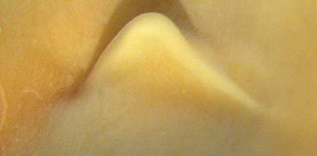
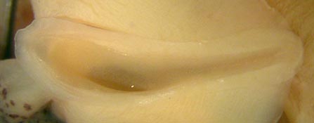
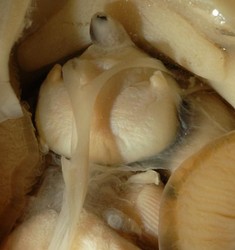
- Funnel locking-apparatus with deep, angled, anterior pit.
Figure. Funnel/mantle locking-apparatus of I. iris, Hancock Seamount, 29°46'30"N, 179°03'36"E, NMNH 817723, left in photographs is anterior. Top - Side-oblique view of the mantle component. Bottom - Frontal view of the funnel component. Photographs by M. Vecchione.
- Mantle (Harman and Seki, 1990)
- Dorsal mantle broadly fused to head (fusion approximately reaches posterior midpoints of eyes).
- Ventral-mantle shield large (ca. 80% of ventral mantle length); extends nearly to anterior margin of eyes; with medial anterior indentation.
- Mantle with pronounced middorsal arch.
- Fins
- Fins with pointed posterior lobes; fins extend to posterior margin of mantle (beyond margin in preserved animals).
- Photophore
- Visceral photophore with strongly elevated papillae each composed of two two distinct tubes.
- Photophore tubes located at lateral edges of photophore. Presumably the tubes enter the photophore between the "lens" and the ink sac but this needs confirmation.
- Pigmentation
- Posterior region of head between edge of secondary eyelid and collor with distinct chromatophore-free band.
 Click on an image to view larger version & data in a new window
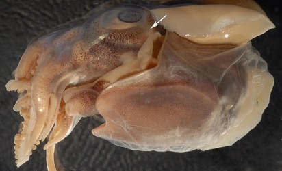
Figure. Ventrolateral view of the posterolateral portion of the head of I. iris, showing chromatophore pigmentation in the region where the lateral funnel adductor attaches. Arrow points to chromatophore-free band. Photograph by R. Young.
- Posterior region of head between edge of secondary eyelid and collor with distinct chromatophore-free band.
- Measurements and counts (Harman and Seki, 1990)
Sex Male Male
Male
Male
Female Female
Female
Female
Mantle length 8.5 11.5 15.5 24.1 9.5
13.0
15.5
28.4
Total length 19.5 31.0 41.0 60.1 24.0 28.5 32.0 46.6 Head width 12.5 14.5 18.0 20.2 13.5 15.0 17.3 17.4 Ventral mantle length 13.0 20.0 24.5 30.0 13.5 20.0 21.5 29.8 Mantle width 7.0 11.0 13.5 16.2 7.5 10.5 11.4 15.0 Shield length 11.5 17.0 20.5 24.8 12.3 17.0 20.9 26.9 Eye diameter 7.5 11.0 13.5 15.7 6.7 10.0 11.5 14.5 Eye lens diameter 3.0 3.0 4.4 4.5 2.5 3.5 2.0 3.4 Fin width 26.5 28.5 37.0 42.5 21.3 29.0 32.1 36.0 Fin length 12.5 16.0 19.0 21.8 12.4 16.5 17.7 18.4 Fin base 6.0 8.0 19.5 12.2 6.2 7.0 8.4 7.7 Tentacle club length 4.5 5.0 6.5 7.1 3.5 6.0 6.0 9.5 Tentacle length 21.0 29.0 26.0 41.5 11.0 35.0 31.0 51.8 Arm I, length 6.0 14.0 19.5 23.2 7.5 9.0 10.3 13.9 Arm II, length
6.0 14.0 19.5 23.3 8.0 9.0 10.4 15.5 Arm III, length
7.5 15.0 22.5 28.2 9.0 10.0 13.8 14.9 Arm IV, length
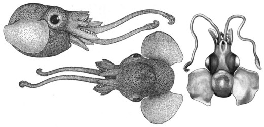
6.0 11.0 15.0 20.1 8.0 9.5 11.4 16.3 - Type illustrations
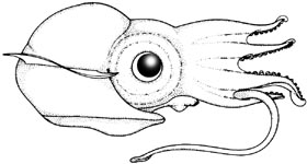


 Go to quick links
Go to quick search
Go to navigation for this section of the ToL site
Go to detailed links for the ToL site