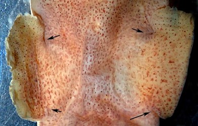

Figure. Dorsal view of New Species B. Photograph by R. Young.
Characteristics
- Arms
- Proximal sucker distinctly largest on all arms.
- Suckers biserial proximally (5-10 suckers on each arm), multiple (3-6) series of suckers on distal 60% (arms IV) to 70% (arms I-III) of arms. Arms IV appear to reach 5-6 series near end of distal 60% of club, 5 series on arm III, 3 or 4 series on arm I and about 4 series on arm II.
- Transition gradual to smaller suckers and more numerous series gradual.
- Low aboral web between arms I, arns I and II, and arms II and III; deep web between arms III and IV.
- Low oral web (about one quarter the depth of the aboral web) between arms III and IV.
 Click on an image to view larger version & data in a new window
Figure. Oral views of suckers of arms of New Species B. Left top - Arm I. Left bottom - Arm II. Right - Arms III (top) and IV. Photographs by R. Young.

Figure. Arm bases of New Species B. Left - Arms II-IV. Right - Arms II-IV. Note the enlarged proximal sucker. Photographs by R. Young.
- Proximal sucker distinctly largest on all arms.
- Tentacles
- Club with approximately 14 suckers in two series on distal portion of club; proximal portion with folded, semilunar keel. Damaged right club appears to have 12 suckers.
- Club with approximately 14 suckers in two series on distal portion of club; proximal portion with folded, semilunar keel. Damaged right club appears to have 12 suckers.
- Buccal crown
- Buccal crown thin, low, delicate and unpigmented. Buccal crown appears to have two ventra supports but connectives to arms IV cannot be seen.
- Buccal crown thin, low, delicate and unpigmented. Buccal crown appears to have two ventra supports but connectives to arms IV cannot be seen.
- Head
- Eyes with corneas.
- Eyes without secondary eyelids.
- Tentacle pocket extends to midline on head posterior to bases of arms IV.
- Anterior eye pore present.
- Anterior eye pocket (part of tentacle pocket) absent.
- Olfactory organ lies with a pocket.
 Click on an image to view larger version & data in a new window
Figure. Side view of the head of New Species B showing the presence of a cornea, absence of a secondary eyelid and a large, circular pit containing the olfactory organ at the posterolateral corner of the head (the pit on the other side of the head was much smaller). Head lightly stained with methylene blue stain. Photograph by R. Young.
- Funnel
- Funnel without lateral funnel adductor muscles.
- Funnel without funnel valve.
- Funnel fused to head except for anterior 2-3 mm and lateral margins.
- Funnel with dorsal and ventral funnel organs.
- Funnle component of the funnel/mantle locking-apparatus with simple, straight groove.
- Mantle component of the funnel/mantle locking-apparatus with simple, straight ridge that extends to the mantle margin.
 Click on an image to view larger version & data in a new window
Figure. Ventral view of an opened funnel of New Species B. Note the absence of the funnel valve. The left arrow points to a ventral pad of the funnel organ and the right arrow points to the dorsal pad. Funnel and adjacent regions stained lightly with methylene blue stain. Photograph by R. Young.
- Funnel without lateral funnel adductor muscles.
- Fins
- Fins broadly separate from eacy other posteriorly.
- Fins with free anterior and posterior lobes; anterior lobe largest.
- Fins rounded laterally.
- Fins broadly separate from eacy other posteriorly.
- Pigmentation
- Numerous reddish brown chromatophores on mantle, head, across nuchal fusion, aboral surfaces of arms (oral surfaces with few, small, scattered chromatopores) and fins (ventral surface of fins without chromatophores on peripherial third).
- Chromatophores more numerous on dorsal surfaces.
- Tentacles with numerous, small and well-separated chromatophores on aboral surface.
- Funnel without chromatophores.
- Numerous reddish brown chromatophores on mantle, head, across nuchal fusion, aboral surfaces of arms (oral surfaces with few, small, scattered chromatopores) and fins (ventral surface of fins without chromatophores on peripherial third).
- Gladius
- Gladius with Y-shape.
- Gladius with central rhachis.
- Vanes diverge posteriorly into large lateral lobes. that Y-shape.
- Gladius extremely thin and fragile.
 Click on an image to view larger version & data in a new window
Figure. Dorsal view of the reconstructed gladius of New Species B. The rhachis is torn and bent at right angles to the gladius (left tip in photograph. The right lobe of the vane tore lose during extraction and has been photographically returned to its natural position. Part of the gladius is missing. Photograph by R. Young.

Figure. Dorsal view of portions of the gladius of New Species B. Left - The anterior tip of the rhachis. Richt - Anterior ends of the vanes. Photographs by R. Young.

Figure. Dorsal view of portions of the gladius of New Species B. Left - Lateral lobe of the left vane. Right - Medial portion of the right vane and the posterior tip of the rhachis. Photographs by R. young.
- Gladius with Y-shape.
- Viscera
- Penis appears to arise from open genital pocket or deep, circular fold in lining of mantle cavity.
- Ventral mantle adductor muscle very thin and weak.
- Large anal flaps present.
 Click on an image to view larger version & data in a new window
Figure. Top - Ventral view of the mantle cavity of New Species B. Arrow points to the penis. Bottom - Spermatangium removed from the penis of New Species B (presumably an aberrant discharge of the spermatophore). Photographs by R. Young.
- Measurements and counts
1Total length of club.Sex Mature male Mantle length 22 mm
Mantle width 14 mm Distance abetween stellate gang. 8 mm Head width 14 mm Head length 8.0 mm Nuchal fusion width 4.6 mm Eye diameter ~ 5.7 mm
Fin length 11.3 mm Fin width (single fin)
7.2 mm Arm I, length 16 mm Arm II, length
18 mm Arm III, length
19 mm Arm IV, length
19 mm Tentacle length 50 mm Club length 2.2 mm1, 1.3 mm2 Web depth, sector A 3.2 Web depth, sector B
3.2 mm Web depth, sector C
3.2 Web depth, sector D 5.3 mm Oral web depth, sector D 1.4 mm
2Length of sucker-bearing region of club.


Figure. Tentacular club of New Species B. Left - Oral view. Right - Aboral view. Photographs by R. Young.


 Go to quick links
Go to quick search
Go to navigation for this section of the ToL site
Go to detailed links for the ToL site