
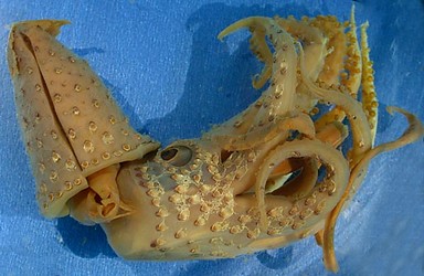
Figure. Ventral view of Histioteuthis eltaninae, holotype, female, preserved, 40°05'S, 149°55'W. Photograph by R. Young.
- Photophores
- Large and small compound photophores intermixed over entire ventral mantle.
- Compound photophores number 18 (17 large, 1 small) on right eyelid (right drawing). Compare with photophores on left eyelid (left drawing).
- Arms IV with 3 longitudinal series on arm base. The upper drawing, to the right, is an enlargement of the arm tip of the lower drawing.
- Terminal group of large, simple photophores on arm I-III absent.
Click on an image to view larger version & data in a new window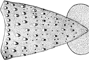
Figure. Ventral view of the mantle of H. eltaninae, holotype, 53 mm ML, female. The small compound photophores are seen as black dots the this drawing. Drawing extracted from Fig. 13a of Voss, 1969.
Click on an image to view larger version & data in a new window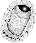
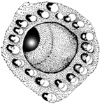
Figure. Lateral views of right and left eyelids of H. eltaninae, holotype. Drawings from Voss, 1969 (Fig. 13d,e).
Click on an image to view larger version & data in a new window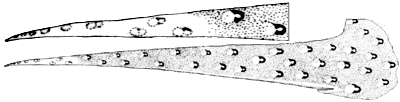
Figure. Ventral view of arm IV of H. eltaninae, holotype. Top - Enlargement of arm IV tip. Bottom - Entire arm IV. Drawing from Voss, 1969 (Fig. 14i).
- Tentacles
- Suckers of manus in 5-6 irregular series.
- Four median suckers on manus enlarged 3-4 times ventral-marginal suckers.
Click on an image to view larger version & data in a new window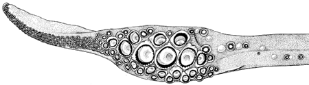
Figure. Oral view of club of H. eltaninae, paratype, 57 mm ML male, 56° 06' S, 71° 14' W. Drawing from Voss, 1969 (Fig. 13b).
- Arms
- Length 100-127% of ML.
- Length 100-127% of ML.
- Sucker dentition
- Largest suckers on manus with rings bearing 36-52 (52 in holotype) low, blunt triangular teeth around entire margin.
- Arm sucker rings with variable number of low, triangular teeth ranging from 7-9 (holotype) confined to distal margin to 20 around entire margin.
Click on an image to view larger version & data in a new window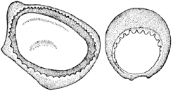
Figure. Suckers of H. eltaninae. Left - Oral view of largest club sucker. Drawing from Voss, 1969 (Fig. 14j). Right - Oral view of large sucker from row 14 of arm II, holotype. Drawing from Voss, 1969 (Fig. 14k).
- Web and buccal crown
- Inner web low to vestigial; outer web absent.
- Buccal crown with 7 supports.
Click on an image to view larger version & data in a new window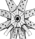
Figure. Oral view of buccal crown of H. eltaninae, holotype. Drawing from Voss, 1969 (Fig. 14l).
- Funnel organ
- Separate low ridge on median of each arm of dorsal pad. Click on an image to view larger version & data in a new window
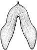
Figure. Ventral view of funnel organ of H. eltaninae, holotype. Drawing from Voss, 1969 (Fig. 14m).
- Separate low ridge on median of each arm of dorsal pad.
- Occipital folds
- One fold present.
- One fold present.
- Beaks
- Ridge on lateral wall of lower beak absent.
- Ridge on lateral wall of lower beak absent.
- Fins
- Length 29-37% of ML; width 50-55% of ML
- Length 29-37% of ML; width 50-55% of ML
- Spermatophores
- Length 6.9-7.3% of ML; sperm mass 2-4% of spermatophore length (SpL), cement body 67-75% of SpL, ejaculatory apparatus 21-26% of SpL and with single loop. Connective complex long.
- Length 6.9-7.3% of ML; sperm mass 2-4% of spermatophore length (SpL), cement body 67-75% of SpL, ejaculatory apparatus 21-26% of SpL and with single loop. Connective complex long.
- Hectocotylus
- Mature male with suckers on basal portions of arms I (and perhaps all arms) slightly enlarged with swollen, fleshy collars. No other hectocotylization known.
Comments
The above data are from Voss (1969) and Voss, et al. (1998).


